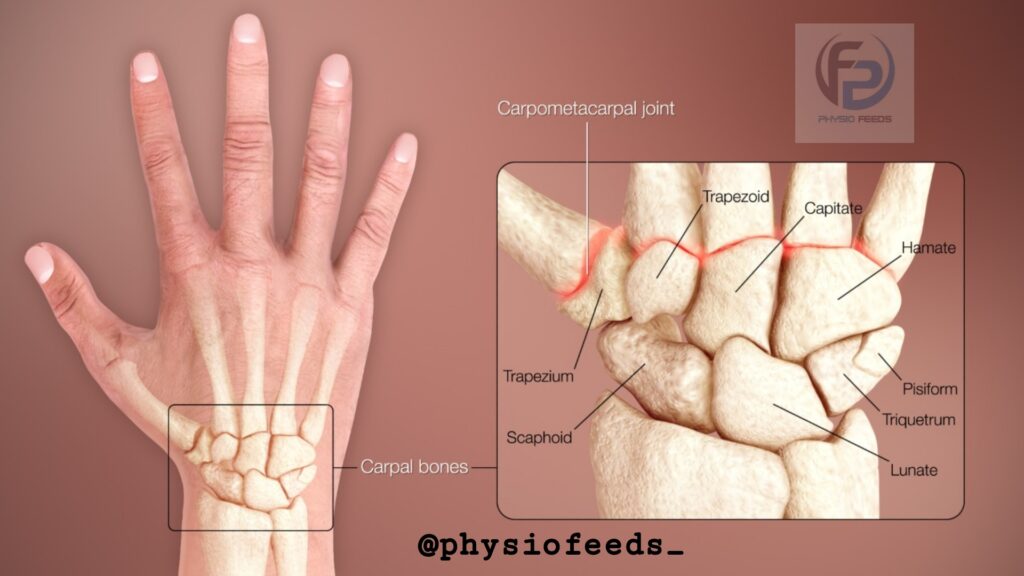
CARPAL BONE ( WRIST BONE)
– The wrist made up of 8 carpal bones (wrist bone) and they are arranged in 2 rows.
1) The proximal row ( from lateral to medial side ) consists :
✓ The scaphoid
✓ The lunate
✓ The triquetral
✓ The pisiform
2) The distal row consists ( in same order )
✓ The trapezium
✓ The trapezoid
✓ The capitate
✓ The hamate
• IDENTIFICATION
1) The scaphoid
– Boat shaped
– Has a tubercle on its lateral side.

2) The lunate
– Half moon shaped or crescentic.

3) The triquetral
– Pyramidal in shape.
– Has an isolated oval facet ( on distal part of palmar surface ).

4) The pisiform
– Pea shaped.
– It has only one oval facet ( on proximal part of its dorsal surface ).

5) The trapezium
– Quadrangular in shape.
– It has a crest and groove anteriorly.
– It has a sellar articular surface distally .

6) The trapezoid
– Resembles the shoe of a baby.

7) The capitate
– Largest carpal bone with a rounded head.

8) The hamate
– Wedge shaped with hook near its base.

• SIDE DETERMINATION
* General points
1) The proximal row of carpal bone (wrist bone)
– Convex proximally
– Concave distally
2) The distal row
– Convex Proximally
– Flat distally
– The palmar and dorsal suface : non articular except for triquetral and pisiform
– Lateral surface of the scaphoid and trapezium : Non Articular
– The medial surface of triquetral , pisiform and hamate : non Articular
3) The Dorsal non-articular surface is always larger than palmar non-articular surface , except for lunate , in which the palmar surface is larger than the dorsal surface .
* Specific points
1) The scaphoid
– Tubercle is directed laterally , forward and downward.

2) The lunate
– A small semilunar articular surface for the scaphoid is on the lateral side.
– A quadrilateral articular surface for triquetral on medial side.

3)The triquetral
– Oval facet for the pisiform lies on distal part of palmar surface
– Medial and dorsal surface are continuous and non-articular.

4) The pisiform
– The oval facet for triquetral lies on proximal part of dorsal surface
– the lateral surface is grooved by ulnar nerve.

5) The trapezium
– The palmar surface has a vertical groove for tendon of Flexor carpi radialis
– The groove is limited laterally by crest of trapezium
– The distal surface bears a sellar concavo-convex articular surface for base of 1 st metacarpal bone.

6) The Trapezoid
– Distal articular surface is bigger than proximal
– Palmar non-articular Surface is prolonged laterally.

7) The capitate
– Dorsomedial angle is distal-most projection from body of the capitate .
– It bears a small facet for 4 th metacarpal bone.

8) The hamate
– The hook projects from distal part of palmar surface and is directed laterally.

• ATTACHMENTS
– There are 4 bony pillars at 4 corners of corpus.
– All the attachments are to these 4 pillars,
1) The tubercle of scaphoid
– The flexor retinaculum
– A few fibres of abductor pollicis longus.
2) The pisiform gives attachment to,
– Flexor carpi ulnaris
– Flexor retinaculum and it’s superficial slip
– Abductor digiti minimi
– Extensor retinaculum
3) The trapezium
– The crest gives origin to Abductor pollicis brevis, flexor pollicis brevis and opponens pollicis ( Thenar eminence ).
– Edge if groove give attachment to 2 layers of flexor retinaculum .
– Lateral surface gives attachment to lateral ligament of wrist joint
– Groove lodges the tendon of Flexor carpi radialis.

4) The hamate
– Tip of hook gives attachment to the flexor retinaculum.
– Medial side of hook gives attachment to flexor digiti minimi and opponens digiti minimi.
• ARTICULATIONS

1) The scaphoid
– Radius, lunate, trapezium ,trapezoid and capitate
2) The lunate
– Radius , scaphoid , capitate , hamate and triquetral
3) The triquetral
– Pisiform , lunate, hamate and articular disc of inferior radioulnar joint
4) The pisiform
– Articulates with triquetral
5) The trapezium
– Scaphoid , 1 st and 2 nd metacarpal and trapezoid
6) The Trapezoid
– Scaphoid , trapezium, 2 nd metacarpal and capitate
7) The capitate
– Scaphoid , lunate, Hamate, 2 nd , 3 rd and 4 th metacarpal and trapezoid.
8) The hamate
– Lunate , triquetral , capitate and 4 th and 5 th metacarpal bones.
• OSSIFICATION
1) Capitate – 2 months
2) Hamate – 3 months
3) Triquetral – 3 years
4) Lunate – 4 years
5) Scaphoid – 5 years
6) Trapezium – 5 years
7) Trapezoid – 5 years
8) Pisiform – 12 years.
• CLINICAL ANATOMY

1) Fracture of scaphoid
– This fracture occurs through the waist at right angles to its long axis
– Fracture caused by fall on outstretched hand or on tip of the fingers
– This causes tenderness and swelling in Anatomical snuff box
– Also causes pain on longitudinal percussion of thumb and index finger
– The importance of fracture lies inits liability to nonunion and avascular necrosis of body of the bone .
– Normally , the scaphoid has 2 nutrient arteries , one entering the palmar surface of the tubercle and other the dorsal surface of the body.
– In about 13% of cases both vessels enter through the tubercle or through distal half of the bone .
– In such cases , fracture may deprive the proximal half of the bone of its blood supply leading to avascular necrosis.

2) Dislocation of lunate
– May be produced by fall on acutely Dorsiflexion of hand with elbow flexion.
– This displaces lunate anteriorly , also leading to carpal tunnel syndrome like features.

THANK YOU
BY PHYSIOFEEDS.
FOLLOW ME IN SOCIAL MEDIA