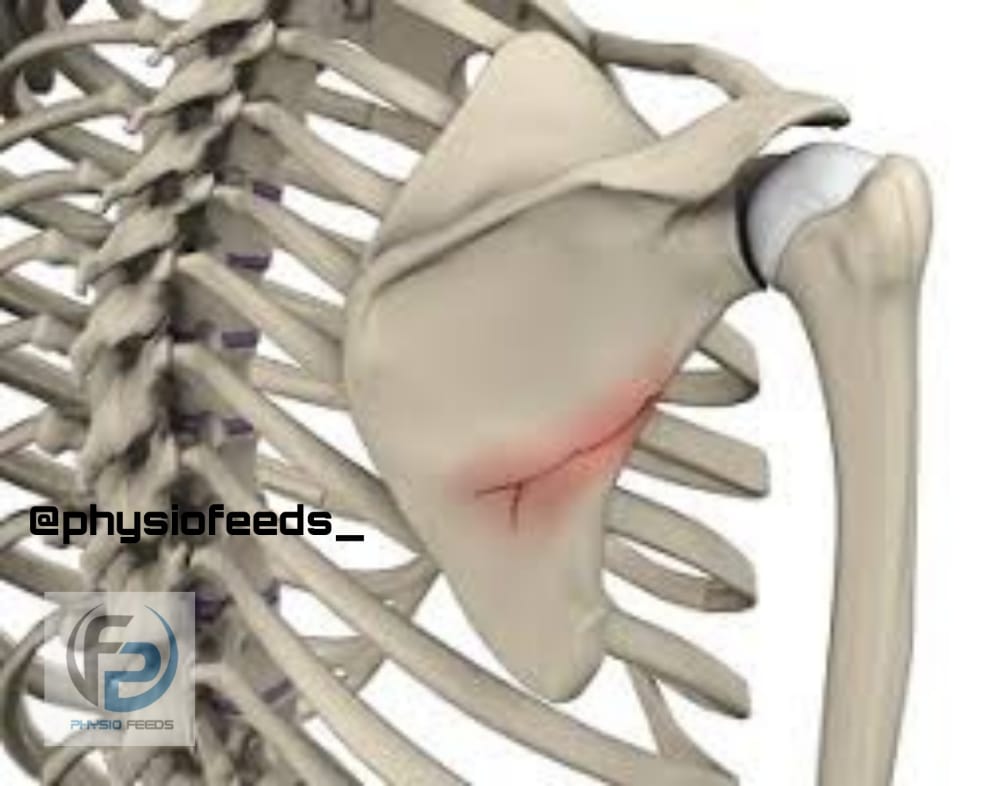
SCAPULA (SHOULDER BLADES)

-Other name of scapula is shoulder blades.
– The scapula is a thin bone.
– It is placed on posterolateral aspect of thoracic cage .
– Scapula has ;

2 surfaces – Costal surface
Dorsal surface
3 borders – Superior border
Lateral border
Medial border
3 processes – superior angle
Inferior angle
Lateral or glenoid angle.
• Side determination

– Lateral or glenoid angle is large and it bears the glenoid cavity.
– Dorsal surface is convex and costal surface is convex.
– Thickest lateral border runs from glenoid cavity above to inferior angle below.
• Features
* Surfaces
1) The costal surface or suprascapular fossa :
– It is concave and is directed medially and forwards.
– Marked by 3 longitudinal ridges.
2) The Dorsal surface :
– Gives attachment to spine of scapula which divides the surface into Supraspinous fossa and infraspinous fossa.

*Borders
1) The Superior border :
– Thin and shorter
– It present suprascapular notch near the root of coracoid process .
2) The lateral border :
– Thick
– It presents infraglenoid tubercle at the upper end .
3) The medial border :
– Thin
– Extends from superior angle to inferior angle.
* Angles
1) The Superior angle
– It is covered by trapezius.
2) The inferior angle
– It is covered by latissimus dorsi
3) The lateral or glenoid angle
– It is broad and bears the glenoid fossa or cavity .
* Processes
1) The spine or spinous process
– It is a triangular plate of bone with 3 borders and 2 surfaces.
– It divides the dorsal surface of scapula into Supraspinous and infraspinous fossae.
– Posterior border is called as the crest of spine
– This crest has upper and lower lips.

2) The acromion
– It has ,
2 borders : medial and lateral
2 surfaces : superior and inferior
And a facet for clavicle.
3) The corocoid
– This process is directed forwards and slightly laterally .
– It is bent and finger-like.
• Attachments

1) Subscapularis : arises form medial two-thirds of the subscapular fossa .
2) Supraspinatus : arises from medial two-thirds of Supraspinous fossa.
3) Infraspinatus : arises from medial two-thirds of infraspinous fossa.
4) The deltoid : arises from lower border of the crest of spine and from the lateral border of the acromion .
5) The trapezius : is inserted into upper border of the crest of spine and into the medial border of the acromion.
6) The serratus anterior : inserted along medial border of costal surface.
7) The long head of bicpes brachii : arises from supraglenoid tubercle.
– The Short head is from lateral part of the tip of corocoid process.
8) The coracobrachialis : arises from medial part of the tip of corocoid process.
9) The pectoralis minor : inserted into the medial border and superior surface of the corocoid process.
10) The long head of triceps brachii : arises from infraglenoid tubercle.
11) Teres minor : arises from upper two-thirds of rough strip on dorsal surface along the lateral border.
12) Teres major : arises from lower one-third of rough strip on dorsal aspect of lateral border.
13) The levator scapular : is inserted along the dorsal aspect of medial border , from superior angle up to the root of spine.
14) The rhomboid minor : inserted into dorsal aspect ( medial border ) opposite to root of spine .
15) The rhomboid major : inserted into dorsal aspect ( medial border ) between root of spine and inferior angle.
16) The inferior belly of omohyoid : arises from upper border near the suprascapular notch.
17) The margin of glenoid cavity : This gives attachment to capsule of shoulder joint and to glenoid labrum.
18) The margin of facet on medial aspect of the acromion : This gives attachment to capsule of Acromioclavicular joint .
19) Coracoacromial ligament :
– attached to the lateral border of acromion process
– attached to medial side of tip of acromion process.
20) The coracohumeral ligament : attached to root of corocoid process.
21) The coracoclavicular ligament : attached to corocoid process .
– Trapezoid part on the superior aspect
– Conoid part near the root.
22) Transverse ligament : bridges across suprascapular notch and converts it into foramen .
– The suprascapular vessels lies above this ligament.
23) The spinoglenoid ligament : bridges the spinoglenoid notch .
– The suprascapular vessels and the nerve passes deep to it.

• Ossification
– The scapula ossification starts from one primary centre and seven secondary centres.
* Appearance of primary centre :
– Near glenoid cavity during 8 th week of development.

* Appearance of secondary centre :
– During First year , th first secondary centre appears in middle of corocoid process .
– It fuses by 15 th year.
* Subcorocoid centre :
– It appears during 10 th year , in the root of corocoid process .
– Fuses by 16 th to 18 th years.
* Other centres
– Two for acromion , One for margin of glenoid cavity ( lower 2/3 rd ) ,One for medial border , One for inferior angle.
– These appears at puberty
– Fuses by 25 th year.
• Clinical anatomy
1) Winging of scapula
– Paralysis of serratus anterior causes ‘scapula winging’ .
– The arm cannot be abducted beyond 90°.
– Medial border of scapula becomes more prominent.

2) The scaphoid scapula
– It is a developmental anomoly.
– Here , the medial border of scapula is concave.