ANATOMY OF HEART
INTRODUCTION

-The heart is a muscular pump which is responsible for blood circulation.
-It is a conical hollow muscular organ situated between the lungs in the middle mediastinum.
-It is enclosed within the pericardium.
-It pumps blood to the various parts of the body to meet their nutritive requirements.
-Heart is placed obliquely behind the body of the sternum and the adjoining parts of costal cartilage. So that one-third of it lies to the right and two-third to left to the medial plane.
•Dimension and weight :
-Heart measures about 12 cm in length and 9 cm in breadth.
-It weighs about 300 g in males , 250 g in females.
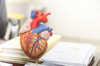
• External features of heart :
✓The human heart has 4 chambers
These are,
-the Right and left atria
-the Right and left ventricles.
✓On the surface of heart they are separated from the ventricles by an atrioventricular groove.
✓ The atria are separated from each other by an interatrial groove and ventricles are separated from each other by an interventricular groove.
✓ The upper part of each atrium has an appendage called the auricle.
✓ The atria separated from the ventricle by a circular atrioventricular sulcus also called as the coronary sulcus.
• Parts of Heart :
1) The apex of heart :
-It is formed by the left ventricle.
-It is directed forwards , downwards and to the left and it is overlapped by the anterior border of left lung.
-Apex is situated in the left 5 th intercostal space, three and half inches lateral to the mid sternal line, just medial to the left midclavicular line.
2) Base of the Heart :
-It is formed by left atrium and by a small part of right atrium.
-It is related to the 5 th to 8 th thoracic vertebrae.
-It is separated from vertebral column by the pericardium.
3) Borders of heart :
– upper border is oblique and is formed by the 2 atria chiefly the left atrium.
-Right border consist entirely of the right atrium.
– Inferior border is made up mostly of the Right ventricle and a small portion of left ventricle.
– Left border is mostly formed by the left ventricle with the auricle of the left atrium.
4) Surface of heart :
– Anterior or sternal costal surface consist of right atrium and the right ventricle and partly by the left ventricle and left auricle.
– Inferior or diaphragmatic surface rest on the central tendon of diaphragm.
It is formed in its left two-third by the left ventricle and in its right one-third by the right ventricle.
– The left surface is formed mostly by the left ventricle and at the upper end by the left auricle.
• Surface marking of Heart :
✓ Right border of heart extends from the lower border of the right 3 rd costal cartilage to the lower border of right 6 th costal cartilage just beyond the right margin of the sternum.
✓ Inferior border passes from the right 6 th costal cartilage to the apex of which is normally in left 5 th intercostal space about three and half inches from the midline.
✓ From the apex of left border extends upwards to lower border of the left 2 nd costal cartilage about 2 cm from the sternal margin.
•Circulation of Blood :
✓The Right Atrium receives deoxygenated blood from the whole body through the superior and inferior vena cava and the coronary sinus.
Atrium gets contract and sends the blood through the right atrioventricular orifice to the right ventricle.
✓The Right Ventricle contracts and propels the blood into the pulmonary trunk to the pulmonary arteries and finally to the lungs where the blood is oxygenated.
The oxygenated blood revert back to the heart through the 4 pulmonary veins and enters the left atrium.
✓The Left Atrium contracts and sends it’s blood through left atrioventricular orifice into left ventricle, which in turn contracts and drives the blood into ascending aorta and it’s ramification.
•Valves of heart :
The valve maintain unidirectional flow of blood and prevents it’s regurgitation in opposite direction.
There are 2 pairs of valves in heart
(1) a pair of semilunar valve
(2) a pair of atrioventricular valve.
√ Right atrioventricular valve is called TRICUSPID VALVE
√ Left atrioventricular valve is called BICUSPID VALVE
•Veins of the Heart :
Veins of the heart falls into 3 groups,
(1) Venae cordis minimas
(2) Anterior cardiac veins
(3) coronary sinus with its normal 5 tributaries.
• 5 Tributaries of coronary sinus ;
(1) Great cardiac vein
(2) Middle cardiac vein
(3) Small cardiac vein
(4) Posterior vein of left ventricle
(5)Oblique vein of left atrium.
•Lymphatics of heart :
-These accompany the coronary arteries and form two trunks.
-The Right trunk ends in brachiocephalic nodes and the Left trunk ends in tracheobrachial lymph nodes at biforcation of trachae.
•Nerve supply :

-Parasympathetic nerve reach the heart via vagus nerve.
These are cardio inhibitory when it is stimulated, they slow down the heart rate.
√ Sympathetic nerves are derived from the upper 3-4 thoracic segments of spinal cord .
These nerves are cardio-acceleratory and when they are stimulated they increase the heart rate and also dilate the coronary arteries
√ Superficial cardiac plexus :
They are situated below the arch of aorta in front of right coronary artery
√Deep cardiac plexus :
They are Situated in front of the bifurcation of trachea and behind the arch of aorta.
• Blood supply of Heart :

Two coronary arteries and their branches supply blood to the heart.
-The right coronary artery and left coronary artery comes from the aortic sinuses at the beginning of ascending aorta and run in the respective atrioventricular grooves.
-There are two principle branches from each;
(1) marginal and posterior interventricular artery from the right coronary artery
(2) the circumflex artery and anterior interventricular artery from the left Coronary artery .
*Branches of the Right coronary artery :
-It has got larger and smaller branches.
The larger branches are ;
(1) Marginal artery
(2) Posterior interventricular artery
The smaller branches are ;
(1) Nodal artery
(2)The right atrial branch
(3) The artery of conus
(4) Terminal branches.
It supplies ,
(1) Right Atrium
(2) Ventricles ;
– A greater part of right ventricle except the part of the area adjoining at the anterior interventricular groove.
– a small part of the left ventricle adjoining at the posterior interventricular groove.
(3) Posterior part of the interventricular septum.
(4)Whole of the conducting system of heart except a part of left branch of the AV bundle.
* Branches of Left coronary artery :
It has got longer and smaller branches.
The longer branches are ;
(1) Anterior interventricular branch
(2) Branch to diaphragmatic surface of the left ventricle.
The smaller branches are ;
(1) Left Atrium branches
(2) Pulmonary branches
(3) Terminal branches.
It supplies ,
(1) Left Atrium
(2) Ventricles ;
– the greater part of left ventricle except the area adjoining at the posterior interventricular groove
– a small part right ventricle adjoining the anterior interventricular groove.
(3) Anterior part of interventricular septum
(4) A part of the left branch of AV bundle.
Pingback: ARTERIAL BLOOD PRESSURE - PHYSIOFEEDS