
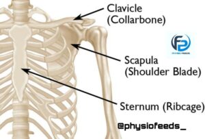
CLAVICAL (THE COLLOR BONE)
-CLAVICAL (THE COLLOR BONE) is a long bone . It is also known as collor bone/ beauty bone.
-It is the only bone in the body that lies horizontally.
-It is a bone of upper extremity.
– It has ;
1) Shaft
2) Two ends – lateral and medial.
• Side determination
√ Lateral End – flat
Medial end – quadrilateral
√ Shaft – slightly curved
* Medial 2/3 rd – convex forwards
* Lateral 1/3 rd – concave forwards.
✓ Clavicle is the only long bone which lies horizontally.
✓ It is the first bone which starts ossifying.
✓ It receives weight of upperlimb via lateral 1/3 rd through coracoclavicular ligament and transmits to axial skeleton via the medial 2/3 rd.
• Features

1) Shaft
– It is divided into lateral 1/3 rd and medial 2/3 rd.
* Lateral 1/3 rd
√ Flattened from above downward
√ 2 borders – Anterior and posterior
Anterior – concave forwards
Posterior – convex backwards
√ 2 Surfaces – Superior and Inferior
Superior – It is subcutaneous.

Inferior – Presents an elevation called conoid tubercle and a ridge called trapezoid ridge.

* Medial 2/3 rd
√ It is rounde
√ 4 surfaces :
Anterior surface – convex forwards
Posterior surface – smooth
Inferior surface – has a rough oval impression at the medial end
Lateral half of the surface has a longitudinal subclavian groove.
There is a nutrient foramen present at the lateral end of the groove.
2) Lateral End / Acromial end
– Flattened from above downwards .
– It has a facet that articulates with acromion process of the scapula and form Acromioclavicular joint.
3) Medial / Sternal end
– It is quadrangular
– Articulates with clavicular notch of manubrium sterni and forms sternoclavicular joint.
• Attachments

1) At Lateral End ;
– The margin of articular surface for its Acromioclavicular joint gives attachment to joint Capsule.
2) At medial end ;
– Margin of articular surface for the sternum gives attachment to ,
(a) Fibrous capsule of sternoclavicular joint
(b) Articular disc ( posterosuperiorly )
(c) Interclavicular ligament ( superiorly )
3) Lateral 1/3 rd of shaft
(a) Anterior border – it gives origin to deltoid .
(b) Posterior border – It provides insertion to trapezius.
(c) Conoid tubercle and trapezoid ridge – Gives attachment to Conoid and trapezoid parts of coracoclavicular ligament.
4) Medial 2/3 rd of shaft
(a) Anterior surface : gives origin to pectoralis major
(b) Half of the superior surface : gives origin to Clavicular head of Sternocleidomastoid muscle .
(c) The oval impression on inferior surface ( at the medial end ) : It gives attachment to costoclavicular ligament.
(d) The subclavian groove : It gives insertion to subclavius muscle
(e) Posterior surface close to medial end : it gives attachment to sternohyoid muscle.
(f) Subclavian vessels and cords of brachial plexus passes towards axilla which lies between inferior surface of clavicle and upper surface of 1st rib .
– The nutrient foramen : transmits a branch of suprascapular artery.
• Ossification of clavicle

– It is the first bone to ossify
– It ossifies in membrane ,except for its medial end.
– Ossification starts from 2 primary centres and 1 secondary centre.
* Two primary centres :
– Appears in shaft
– Between 5th and 6 th week of intrauterine life.
– And fuse about the 45th day..
* The secondary centre
– Appears during 15-17 years
– Fuses with shaft during 21-22 years.
• Clinical anatomy
1) Clavicle fracture

– Clavicle is most commanly fractured.
– This fracture can occur by falling on outstretched hand .
– Most common site : junction between two curvatures of the bone.

2) Cleidocranial dysostosis
– Congenital absence of clavicle or imperfectly developed clavicle in a disease is called as Cleidocranial dysostosis.
